Shining a light on optical imaging
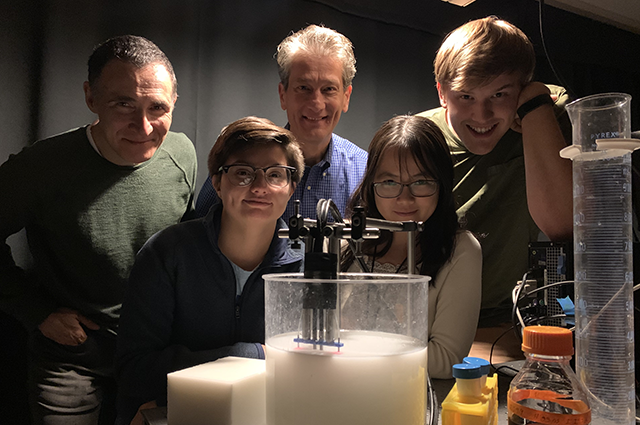
In the Diffuse Optical Imaging of Tissue (DOIT) Lab at Tufts University, Professor Sergio Fantini and collaborators – including Research Assistant Professor Angelo Sassaroli and Ph.D. candidates Giles Blaney, Thao Pham, and Cristianne Fernandez – study biological tissue at a macroscopic scale. When medical professionals and researchers need to scan patients’ tissue or organs, the availability of modern imaging techniques means that an invasive biopsy isn’t always necessary. One of those imaging techniques is near‐infrared spectroscopy (NIRS), which sheds light on the dynamics of blood flow and the oxygenation of tissue. In the DOIT Lab, Fantini and colleagues recently published three papers on a new approach to NIRS that offers better sensitivity and more targeted study of deeper biological tissue.
When areas to be studied lie beneath superficial tissue layers – like the brain under scalp and skull or skeletal muscle under skin and fatty tissue – those tissue layers can confound measurements. Instruments pick up blood flow changes in the scalp, for instance, while taking brain measurements. When it comes to NIRS technology and the diffuse reflection of light, the region of sensitivity is shaped like a banana and is at its most sensitive closest to the source-and-detector pairing that creates that region – instead of being most sensitive deeper in the tissue.
Currently, researchers strive to measure and quantify confounding effects from superficial tissue, so that, for example, the effects of blood flow in the scalp can be identified and removed from consideration in optical imaging of the brain. That work has been done with variable success, so Fantini and the DOIT Lab proposed a novel approach: suppressing the sensitivity of optical signals to these confounds. They developed a new arrangement with a dual-slope approach that calls for at least two sources and two detectors laid out in specially-configured arrays. Their proposed approach would allow researchers to study localized points deeper in tissue, as opposed to the current larger, banana-shaped region that also captures confounding data from nearby superficial tissue.
The Tufts researchers’ results, in studying the new dual-slope approach, demonstrated major advances over more traditional single-distance or single-slope data, allowing for greater depth sensitivity in NIRS imaging. The DOIT Lab continues to develop new advances in diffuse near-infrared spectroscopy and the non-invasive imaging of biological tissue, with a provisional patent filed and further research to come.
Learn more:
Angelo Sassaroli, Giles Blaney, and Sergio Fantini, “Dual-slope method for enhanced depth sensitivity in diffuse optical spectroscopy,” Journal of the Optical Society of America A 36, 1743-1761 (2019).
Giles Blaney, Angelo Sassaroli, Thao Pham, Cristianne Fernandez, and Sergio Fantini, “Phase dual slopes in frequency-domain near-infrared spectroscopy for enhanced sensitivity to brain tissue: First applications to human subjects,” Journal of Biophotonics, c201960018 (2019); https://doi.org/10.1002/jbio.201960018.
Sergio Fantini, Giles Blaney, and Angelo Sassaroli, “Transformational change in the field of diffuse optics: From going bananas to going nuts,” Journal of Innovative Optical Health Sciences. 1930013 (2019); https://doi.org/10.1142/S1793545819300131.
Department:
Biomedical Engineering