About the Center

The Tufts Advanced Microscopic Imaging Center (TAMIC) offers a wide array of optical and spectral quantitative imaging techniques, for the chemical and structural characterization of materials at submicron scales. Our staff is particularly experienced in confocal imaging and two-photon imaging of biological specimens and engineered tissues. Laser excitation and photodetection schemes are optimized for label-free functional imaging of live cells and tissues, such as:
- Cellular metabolism and mitochondrial redox state (Two-photon excited fluorescence, TPEF)
- Lipid metabolism and neuronal myelination (Coherent anti-Stokes Raman spectroscopy, CARS)
- Collagen fiber organization (Second harmonic generation, SHG)
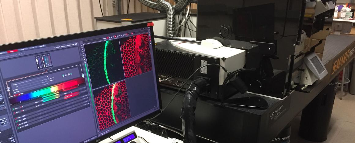
The centerpiece of the imaging center is a pair of Leica SP8 confocal/multiphoton microscopes which can provide:
- Long-term, label-free imaging of live cells and tissues
- 3D imaging with submicron spatial resolution at depths < 200µm
- Multimodal microscopy (confocal reflectance, fluorescence, TPEF, SHG, CARS, FLIM, DIC)
- Excitation lasers at 405, 448, 514, 552, 638 (CW) and 680-1300nm (pulsed)
- NIR multiphoton excitation for reduced photodamage and deeper penetration
- Multicolor confocal imaging with 3 independent, continuously tunable photodetectors
- Fluorescence lifetime imaging (FLIM) for environmentally-sensitive molecular probing
- Macroscopic imaging at submicron scales (image stitching)
- Extremely sensitive photon counting detectors (Leica HyD)
- Video rate scanning (for 256x256 images)
- Motorized stage for autofocus and automated microplate imaging
TAMIC is supported by a grant from the National Institutes of Health Division of Program Coordination, Planning, and Strategic Initiatives' Office of Research Infrastructure Programs, and from the National Science Foundation Major Instrumentation Research grant.