Research
Optical assessment of tissue function for tissue engineering and regenerative medicine applications
The two main tissue aspects that we aim to characterize optically include cellular metabolism and collagen organization. Cell metabolism plays a central role in the regulation of normal development, the function and the death of a cell. There are two co-enzymes involved in key metabolic pathways, NADH and FAD, that naturally absorb light in the visible range of the spectrum and re-emit it at slightly longer wavelengths. These signals can be detected and quantified to assess the metabolic state of the cells. This information in turn can be used to assess normal or diseased cell development.
Biochemical, morphological and organizational changes associated with very early cancer development
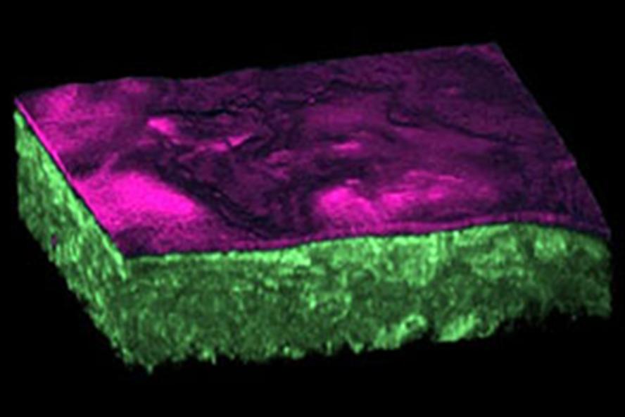
Epithelia constitute one of the four basic animal tissue types. They cover or line organ surfaces and cavities and serve multiple functions, including protection, secretion and absorption. Non-invasive approaches to assess epithelial cancerous transformation could greatly impact clinical diagnostics, as the majority of cancers are of epithelial origin. We have employed Engineered Epithelial Tissue Equivalents, constructed by either primary human foreskin keratinocytes (i.e. normal human epithelial cells) or immortalized human foreskin keratinocytes expressing the full-length genome of human papilloma virus 16 (HPV16), as a biological model to study HPV-induced epithelial pre-cancer. These are tissues that we can grow in the lab and they look very similar to normal human tissues (Fig. 2). In fact, these are the types of tissues that are already commercially available for transplantation in patients with severe skin injuries, as a result of burns or ulcers, for example.
Non-destructive characterization of collagen in normal and diseased tissue development
We have been developing imaging methods and analysis software to characterize collagen fiber organization for different biomedical applications (Bayan et al., 2009; Quinn and Georgakoudi, 2013). By utilizing non-invasive imaging techniques (e.g. two photon excited fluorescence, second harmonic generation, and confocal reflectance microscopy) and developing quantitative outcomes related to tissue composition and organization, we can provide unique insight into structural and mechanical changes during dynamic events that occur as tissues develop normally or when diseases, such as cancer, arise.
Bayan C, Levitt J, Miller E, Kaplan D, Georgakoudi I. Fully-automated, quantitative, non-invasive assessment of collagen fiber content and organization in thick collagen gels. Journal of Applied Physics 2009, 105:102042.
Quinn KP, Georgakoudi I. Rapid quantification of pixel-wise fiber orientation data in micrographs. Journal of Biomedical Optics 2013; 18 (4): 046003

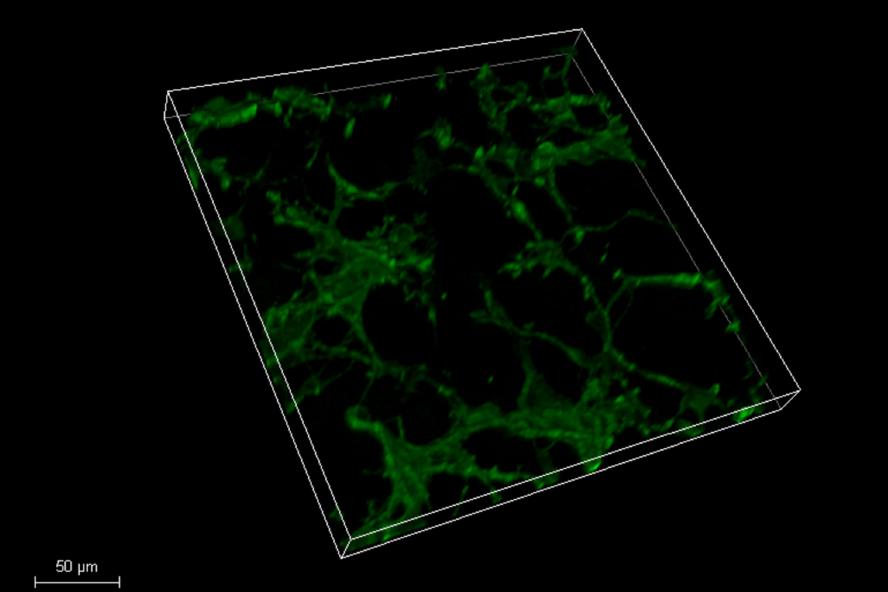
FIG. 1
3D Fluorescence image of 1.5 week old cerebellar neurons stained with ß-3-tubulin. (Photo: Will Collins)

FIG. 2
3D reconstruction of Engineered Epithelial Tissue Equivalent constructed by primary human
foreskin keratinocytes. Cellular layers are shown in green and the keratinized layer is shown in
magenta. (Photo: Dimitra Pouli)

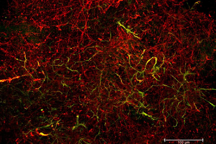
FIG. 3
3D fluorescence image of a co-culture of murine fetal cortical neurons and astrocytes in a silk
scaffold, incubated for 1 week and immunostained against the main neuronal Tuj1 and astrocytic
GFAP markers. (Photo: Olga Liaudanskaya)

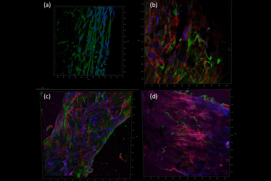
FIG. 4
3D fluorescence images of:
(a) stained muscle cells; (b-d) co-cultures of nerve stem cells (green) and muscle cells (pink, red). (Photo: Tom Dixon)