Research/Areas of Interest
label-free high resolution tissue imaging, non-linear microscopy, metabolic imaging, matrix characterization, in vivo flow cytometry, cancer detection, osteoarthritis, neurodegenerative diseases
Education
- B.A., Dartmouth College, Hanover, United States, 1993
- M.Sc., University of Rochester, Rochester, United States, 1996
- Ph.D., University of Rochester, Rochester, United States, 1999
Biography
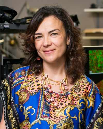
Irene Georgakoudi has been working on the use of lasers for therapeutic and diagnostic applications since her undergraduate years. She started as a physicist at Dartmouth College and continued her graduate studies in biophysics at the University of Rochester. Her interests in spectroscopy and spectroscopic imaging using endogenous sources of contrast were founded during her postdoctoral years at the MIT Spectroscopy Lab. After working on the development of fluorescence-based in vivo flow cytometry while an instructor at the Wellman Laboratories for Photomedicine at Massachusetts General Hospital/Harvard Medical School, Georgakoudi joined Tufts in 2004. She is the author of several patents on the development and use of spectroscopy and imaging to characterize tissues or to detect specific populations of cells, and has published numerous peer-reviewed manuscripts, review articles, and book chapters on these topics. She is the recipient of a Claflin Distinguished Scholar, an NSF Career Award, and an American Cancer Society Research Scholar award. She has served on the Board of Directors of the Optical Society of America and she is an Associate Editor for the journal Optica. She is a fellow of the American Institute for Medical and Biological Engineering, Optica (previously the Optical Society of America) and the International Society for Optics and Photonics (SPIE).
Irene Georgakoudi's lab is interested in the development of new methods to assess different aspects of the normal or diseased development of human tissues that rely on light interactions with molecules that are naturally present in our cells and tissues. They are thus non-invasive (or minimally invasive) and do not require exogenous contrast agents. The goal is to generate high resolution (microscopic) images of tissues that provide not only morphological, but also functional information about the tissue by bringing the microscope to the patient, instead of using current approaches that rely on excising the tissue and processing it before looking at it under a microscope. These optical methods obviate the need for a biopsy and enable the study of the same specimen dynamically over time. They do not interfere with the physiology of the subject and they do not suffer from artifacts. Our expectation is that these methods will yield critical new insights regarding how conditions such as cancer, osteoarthritis, cardiovascular and neurodegenerative diseases develop and will provide a new paradigm for disease detection.
Irene Georgakoudi's lab is interested in the development of new methods to assess different aspects of the normal or diseased development of human tissues that rely on light interactions with molecules that are naturally present in our cells and tissues. They are thus non-invasive (or minimally invasive) and do not require exogenous contrast agents. The goal is to generate high resolution (microscopic) images of tissues that provide not only morphological, but also functional information about the tissue by bringing the microscope to the patient, instead of using current approaches that rely on excising the tissue and processing it before looking at it under a microscope. These optical methods obviate the need for a biopsy and enable the study of the same specimen dynamically over time. They do not interfere with the physiology of the subject and they do not suffer from artifacts. Our expectation is that these methods will yield critical new insights regarding how conditions such as cancer, osteoarthritis, cardiovascular and neurodegenerative diseases develop and will provide a new paradigm for disease detection.