Modeling traumatic brain injuries

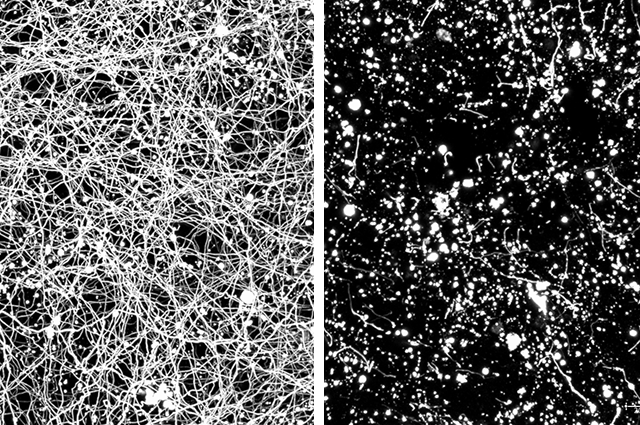
Reaching a better understanding of the long-term effects of traumatic brain injury (TBI), which include neurodegeneration and mental illness, could be life-changing for TBI survivors. A team of researchers from the Tufts University Department of Biomedical Engineering and from Massachusetts General Hospital have developed a 3D contusion model – using mouse cortical neurons grown on a silk scaffold embedded in collagen – and compared it to an in vivo model to provide critical benchmarking for the 3D model’s use in studying traumatic brain injury.
The researchers subjected the 3D brain-like culture system to controlled cortical impact (CCI), a method of studying brain trauma that makes use of a controlled piston to induce injury. They ultimately found that their system mirrored a number of responses to CCI in the in vivo model, giving further credence to the model’s potential in studying the effects of traumatic brain injury and the development of new treatments and therapeutics.
The Tufts team, largely affiliated with the Department of Biomedical Engineering and the Initiative for Neural Science, Disease & Engineering, included postdoctoral scholar Volha Liaudanskaya (first author), PhD student Craig Mizzoni, former postdoctoral Nicolas Rouleau (now an assistant professor at Algoma University), alum Alexander Berk (E18), senior Julia Turner (A20), Professor Irene Georgakoudi, Research Associate Professor Thomas Nieland, and Stern Family Professor in Engineering and Department Chair David Kaplan (corresponding author). They collaborated with colleagues Joon Yong Chung, Limin Wu, and Michael Whalen of the Neuroscience Center at Massachusetts Genera Hospital.
Read "Modeling controlled cortical impact injury in three-dimensional brain-like tissue cultures" in Advanced Healthcare Materials.
Department:
Biomedical Engineering